Cardiac Muscle Cell
Heart Muscle Cell
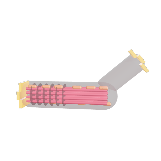
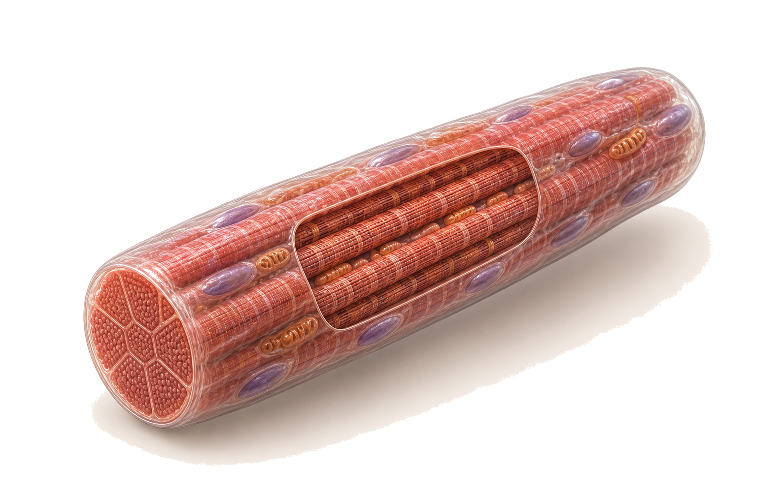
About the Cardiac Muscle Cell
Heart Muscle Cell
Wall of the heart
Cardiac muscle cells form the heart wall and contract in rhythm without conscious control.
Clinical and real-world context
Heart muscle cells depend on constant oxygen. When blood flow stops they die quickly, which is what happens in a heart attack.
4 structures to explore
Intercalated Disc
The electrical junction
These stepped junctions lock neighbouring cells together and let electrical signals pass directly, so the heart contracts as one.
Intercalated discs are found only in cardiac muscle.
Myofibrils
Striped contractile threads
Like skeletal muscle, cardiac cells contain repeating sarcomeres of actin and myosin, but they are shorter and often branch.
The heart beats about 100,000 times a day.
Mitochondria
Tireless energy supply
Cardiac cells are packed with mitochondria because the heart can never rest, and they rely almost entirely on aerobic energy.
Mitochondria make up around 30 percent of a heart muscle cell's volume.
Central Nucleus
Usually one
Unlike skeletal fibres, which have many nuclei at the edge, a cardiac cell usually has one nucleus in the middle.
Adult heart muscle cells divide very rarely.