Chlamydomonas
Single-Celled Alga
About the Chlamydomonas
Single-Celled Alga
Ponds and damp soil
Chlamydomonas swims toward light, using photosynthesis to make food.
Clinical and real-world context
This swimming alga is a model for photosynthesis and for the cilia and flagella that, in humans, are linked to ciliopathies.
4 structures to explore
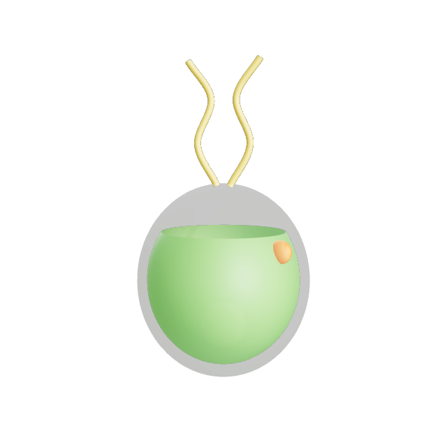
Cup-shaped Chloroplast
One big solar panel
A single large chloroplast wraps around the cell like a cup. A dense pyrenoid inside concentrates the enzymes that fix carbon.
The cup shape is why the cell looks like a green ball with an opening toward its flagella.
Eyespot
The light sensor
The eyespot, with light-sensitive proteins, lets the alga steer toward light that is good for photosynthesis.
The same family of light-sensing proteins is used in optogenetics.
Flagella
The paired oars
Two flagella beat together like breaststroke arms, pulling the cell through water.
These flagella share their structure with human cilia and sperm tails.
Nucleus
Inside the cup
The nucleus sits in the middle of the cell, tucked inside the chloroplast cup.
Chlamydomonas has a haploid genome, so mutations show up directly.